In vivo, real-time, multicolor lymph node imaging based on quantum dots
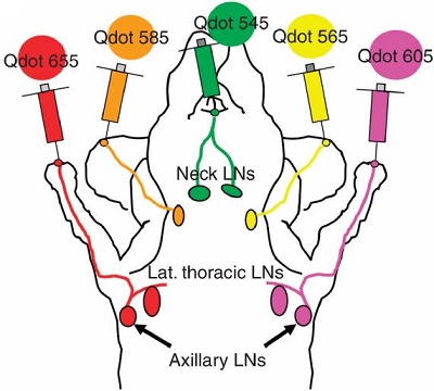
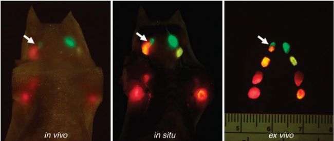
The quantum brightness of Quantum dots (QDs) is very high, and the emission spectrum is narrow and symmetrical. The half-width is less than 30 nm, which can achieve multi-color excitation of a single wavelength, and the mutual interference between multiple emitted light is small. Simultaneous imaging observation of five different colors is possible in the visible range. NIH researcher Kobayashi H et al. subcutaneously injected five different emission wavelength quantum dots (carboxyl-QD 545/565/585/605/655nm) into different parts of mice (Fig. 1), and multicolor fluorescence imaging 5 minutes after injection. Observed, in vivo, real-time imaging of the neck and axillary lymph nodes (Figure 2). In addition, observations and comparisons were made on days 1, 2, and 7 after injection, respectively, and it was found that fluorescence decay was observed after 1 day of injection, possibly due to rapid elution of quantum dots; however, until 7 days after injection, there was almost no subsequent fluorescence. Lost. Although there is a certain loss of fluorescence signal during the above observation process, the emission of all lymph nodes is clear and bright, which can meet the requirements of visual observation and image acquisition.
At present, most lymphatic fluorescence imaging uses near-infrared fluorescence, making full use of the good penetration of near-infrared fluorescence and low auto-fluorescence interference. However, near-infrared fluorescence is invisible to the naked eye, and special acquisition equipment is required. In multi-color imaging, spectral analysis is required to decompose the mixed fluorescence, and the process takes a long time and further post-processing, and cannot be observed in real time. The subject uses the advantages of high fluorescence brightness of quantum dots to achieve multi-color real-time imaging with visual observation, which is of great significance for guiding preclinical and clinical surgical treatment. For example, real-time assisted sentinel lymph node detection analysis facilitates direct, real-time observation of sentinel lymph node resection before surgery.

Figure 1 Schematic diagram of the quantum dot injection site

Figure 2 Lymph node real-time imaging observation
Source of the document:
Kosaka N, Ogawa M, Sato N, Choyke PL, Kobayashi H. In vivo real-time, multicolor, quantum dot lymphatic imaging. J Invest Dermatol. 2009; 129(12): 2818-22.
Microscope for Biology,Bio Lab Microscope,Professional Biological Microscope,Microscopes For Biology
Ningbo ProWay Optics & Electronics Co., Ltd. , https://www.proway-microtech.com