The application of digital microscope in classical sculpture
In the impression of everyone, the ancient marble sculptures are all white. But is this really true? Scientists at the Copenhagen Painted Network (CPN) showcased Greek and Roman sculptures with luxurious decorations and gorgeous colors. With surgical microscopes and digital microscopes, the Heritage Protection Commissioner can detect subtle traces of paint pigments and restore the color feast of ancient times. The Heritage Protection Commissioner used surgical microscopes to record the traces of the Greek marble portraits of the Greek period in 100 BC.
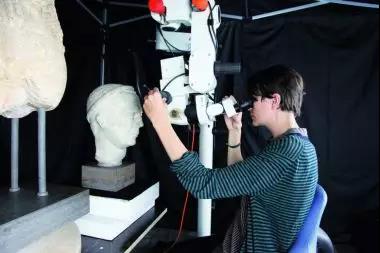
Image source: New Carlsberg Art Museum
In many of the ancient sculptures of the global museums, we have relatively in-depth research on painted sculptures. The Copenhagen Paint Network is an interdisciplinary research team that aims to study the sculptures of the New Carlsberg Art Museum and record the traces of color. The research task is very urgent, because these color traces are gradually disappearing.
Gorgeous colors give life to the restored lion statue.
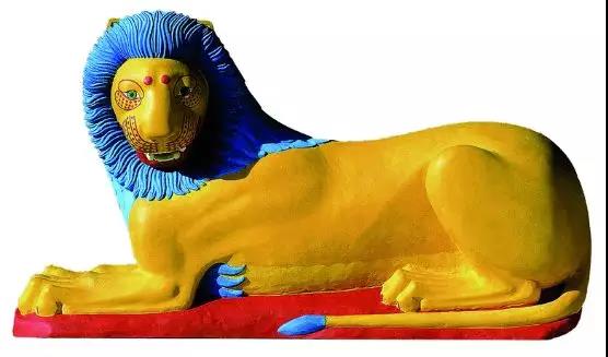
Restorer: V. Brinkmann and U. Koch-Brinkmann
Image source: New Carlsberg Art Museum
Since some of the pigment residue on the sculpture is very rare, making it impossible to extract the sample, the microscope became the most important tool for the Heritage Protection Commissioner Maria Louise Sargent.
“The digital microscope has great flexibility,†said the Heritage Protection Commissioner.
These sculptures are up to two meters high, and our scanning work must be accurate to every centimeter. In addition, the digital microscope can achieve a magnification of 160x. As a result, color pigments and original pigment residues are no longer just traces of traces. Now, we can more easily detect and analyze these clues.
With a digital microscope, we can record videos and images and present them on the monitor during the discussion. After detecting the area of ​​the monitor with pigment residue, the microscope will save the digital image and form a record.
The right eye image of the portrait of a Greek figure obtained with a surgical microscope, the eyelashes and other colored details are clearly visible.
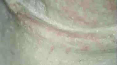
Image source: New Carlsberg Art Museum
Images of the Sphinx's traces of blue pigment obtained with a surgical microscope were unidentified.

Image source: New Carlsberg Art Museum
In the process of searching for traces using a surgical microscope, Sargent found extremely small amounts of blue pigment on the Greek limestone figure in 580 BC.
In the cooperation with the British Museum, CPN is listed as an external research partner to carry out research work.
“Our colleagues at the British Museum have developed a non-invasive method of using UV photography to detect Egyptian blue. We hope to get more information through this technology,†Østergaard said optimistically.
Relics Protection Commissioner Rikke Hoberg used digital microscopes to record the traces of Syrian sculptures.
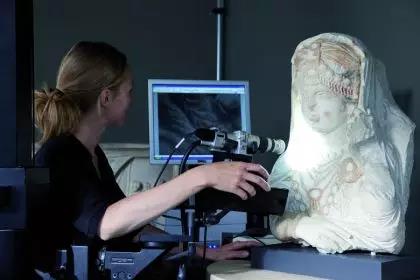
Image source: New Carlsberg Art Museum
Traces of red, ochre and black pigments can be seen through a digital microscope.
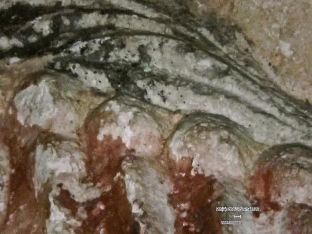
Image source: New Carlsberg Art Museum
Currently, Maria Louise Sargent is working on the beauty of Palmyra, a limestone sculpture dating back to Palmyra, Syria, around 190–210 BC. The harder job is to accurately identify these environmental pollutions. Or traces of erosive agents.
After all, these ancient painters used materials such as ochre and stone as the color of the earth. So which part is the pigment, and which part is only taken from the local soil?
This limestone sculpture is from Pamela, Syria, 190–210 BC, with red jewels on the sculpture.

Image source: New Carlsberg Art Museum
By observing the statues of Greek figures with the naked eye, you can see the traces of various colors on the eyelashes and lips.
Østergaard is looking forward to a truly important discovery: “The detection of this object may help to answer the question of whether the skin of an ancient figure statue has been painted. So far, we have only one evidence from the classical period and the Greek period. Prove that the character's body is painted with skin color pigments."
The portrait of Caligula, the ruler of the Roman Empire during the 37-41 AD, was one of the first Roman statues to be restored using painting.

Image source: New Carlsberg Art Museum
What is the appearance of Caligula? This restoration was for the first exhibition of Bunte Götter in 2003.

Restorers: V. Brinkmann, U. Koch-Brinkmann and JSØstergaard.
Image source: New Carlsberg Art Museum
The Kaliganula restoration map of a later period was completed by the same team on the basis of the same research content, and was more speculative in its higher level of naturalism.
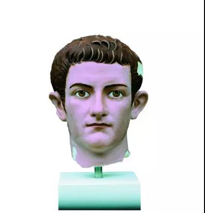
Restorers: V. Brinkmann, U. Koch-Brinkmann and JSØstergaard.
Image source: Stiftung Archäologie, München
Although CPN has achieved some notable research results, the research on painting is still in its infancy.
“Even if we can identify the colors of ancient sculptures, we still don’t know their true face,†Østergaard pointed out the current work problems. “So, we need to understand more details about the craftsmanship and the aesthetics of ancient painted art.â€
About Leica Microsystems Leica Microsystems
Leica Microsystems is a global leader in microscopy and analytical science instruments and is headquartered in Wetzlar, Germany. It mainly provides professional scientific instruments such as research-grade microscopes in the field of microstructure and nanostructure analysis. Since the establishment of the company in the 19th century, Leica has been widely recognized by the industry for its quest for optical imaging and its innovative spirit of continuous improvement. Leica is a global leader in multiple microscopy in composite microscopes, stereo microscopes, digital microscopy systems, laser confocal scanning microscopy systems, electron microscopy sample preparation and medical surgical microscopy.
Leica Microsystems has seven product development and production sites around the world and has service support centers in more than 20 countries. Leica has regional branches or sales branches in more than 100 countries around the world, and has established a comprehensive dealer service network system throughout the world.

Fingerprint Safe Box,Residential Safe,Biometric Safe Box,Steel Safe Box
Hebei Yingbo Safe Boxes Co.,Ltd , https://www.ybsafebox.com