Effective adherent culture of human hPSC using non-coating method of laminin
In 2016, the Miyazaki team at Kyoto University found that during human hPSC subculture, laminin iMatrix-511 can be added to the cell suspension, which eliminates the pre-coating process on the culture dish. Prior to this, it was recognized that it was necessary to spend several hours pre-coating the culture dish to enhance cell adhesion. In addition, the use of iMatrix-511 by non-coating method can reduce the amount of extracellular matrix used, but the efficacy is the same as when the pre-coating method is applied to the culture dish. Finally, the team also confirmed that the addition of iMatrix-511 also supports the long-term maintenance of hPSC single cell passage. By using the non-coated iMatrix-511, hPSC amplification can be made more efficient, less costly and less time- and labor-saving.
Experimental Method <br> hPSC was plated in a medium containing 10 μM Y-27632 and a culture substrate. For the general culture using the iMatrix-511 non-coating method, the concentration of iMatrix-511 was 0.25 μg/cm 2 . For example, in a single well without a coated 6-well plate, 2 mL of a cell suspension containing 5 μL of 0.5 mg/mL iMatrix-511 mother liquor was added for cell culture. After inoculation, hESC H9 cell line and hiPSC (human induced pluripotent stem cell) 253G1 cell line were cultured in TeSR-E8 and StemFit AK03 medium. The cells were treated with a cell digest of 0.5 x trypsin and 5 mM EDTA/DPBS for 4 min every 4 minutes for passage. The volume of the medium was 200 μL/cm 2 .
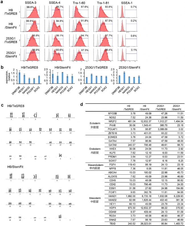
Characteristics of hPSCs grown under non-coated methods <br> The experimental team evaluated whether the long-term culture of hPSCs under non-coated methods was normal through a series of methods. Using the non-encapsulation method of iMatrix-511, hPSC can be serially passaged and passed through more than 10 times. Similar to the cells cultured under the pre-coating method, the hPSC cultured under the non-coated method proliferated well and maintained normal cell morphology. In addition, they highly expressed the representative undifferentiated cell surface markers SSEA-3, SSEA-4, Tra-1-60 and Tra-1-81, and did not express SSEA-1, indicating their continued undifferentiated state. Similar to cells grown under conventional pre-coating (Fig. 1a). The team performed qPCR analysis on hPSCs grown by non-coated methods, and also demonstrated that hPSCs are in an undifferentiated state. The expression levels of the undifferentiated marker genes were similar in comparison to the pre-coating method (Fig. 1b), indicating that there was almost no difference in the undifferentiated state of hPSCs grown in the non-coated and pre-coated methods. Karyotype analysis showed that the non-coating method did not affect the stability of hPSC (Fig. 1c, supplementary Fig. 3). These results indicate that the non-coated method maintains the undifferentiated state of hPSC even after continuous subculture. Finally, the team evaluated the potential of hPSCs grown in non-coated methods to differentiate into different cell lines. hPSC can form embryoid bodies normally. By comparing with the expression of the gene before induction, the expression of the marker genes specific for the three germ layers by the embryoid bodies was highly elevated, indicating that they maintained the differentiation potential (Fig. 1d). Therefore, the team concluded that hPSCs grown under non-coated methods can maintain their pluripotency, and that non-encapsulation can be applied to the daily subculture of hPSC .
 Figure 1. Characteristics of hPSCs grown under non-coated methods . (a) Flow cytometric analysis of representative undifferentiated markers. The gray shaded area on the way was a negative control. (b) qPCR analysis of undifferentiated marker genes. The figure shows the relative expression of 29 passages of hPSC grown under non-coated method and 10 passages of hPSC grown under pre-coating. (c) G band normal karyotype analysis. The hESC H9 cell line has a normal karyotype (46, XX). (d) qPCR analysis of marker genes for embryoid body differentiation. hPSCs were cultured under non-coating and then induced into embryoid bodies over 14 days. By analyzing a single set of data detected by the TaqMan hPSC Scorecard sequence, the expression of a representative differentiation marker gene was compared with the expression of the gene before embryoid formation, and the results indicated that the expression of the genome as shown in the figure was more than twice as high. increase. Data were collected from hPSCs grown for 10 generations under non-coated methods.
in conclusion
iMatrix-511 is effective as a medium supplement and can effectively support cell adhesion when added, but it can be achieved without pre-coating. Unlike other matrix proteins, iMatrix-511 supports cell adhesion and long-term expansion at much lower concentrations than the pre-coating method. This new approach to using iMatrix-511 can reduce the cost and time of hPSC maintenance .
Experimental Method <br> hPSC was plated in a medium containing 10 μM Y-27632 and a culture substrate. For the general culture using the iMatrix-511 non-coating method, the concentration of iMatrix-511 was 0.25 μg/cm 2 . For example, in a single well without a coated 6-well plate, 2 mL of a cell suspension containing 5 μL of 0.5 mg/mL iMatrix-511 mother liquor was added for cell culture. After inoculation, hESC H9 cell line and hiPSC (human induced pluripotent stem cell) 253G1 cell line were cultured in TeSR-E8 and StemFit AK03 medium. The cells were treated with a cell digest of 0.5 x trypsin and 5 mM EDTA/DPBS for 4 min every 4 minutes for passage. The volume of the medium was 200 μL/cm 2 .
Characteristics of hPSCs grown under non-coated methods <br> The experimental team evaluated whether the long-term culture of hPSCs under non-coated methods was normal through a series of methods. Using the non-encapsulation method of iMatrix-511, hPSC can be serially passaged and passed through more than 10 times. Similar to the cells cultured under the pre-coating method, the hPSC cultured under the non-coated method proliferated well and maintained normal cell morphology. In addition, they highly expressed the representative undifferentiated cell surface markers SSEA-3, SSEA-4, Tra-1-60 and Tra-1-81, and did not express SSEA-1, indicating their continued undifferentiated state. Similar to cells grown under conventional pre-coating (Fig. 1a). The team performed qPCR analysis on hPSCs grown by non-coated methods, and also demonstrated that hPSCs are in an undifferentiated state. The expression levels of the undifferentiated marker genes were similar in comparison to the pre-coating method (Fig. 1b), indicating that there was almost no difference in the undifferentiated state of hPSCs grown in the non-coated and pre-coated methods. Karyotype analysis showed that the non-coating method did not affect the stability of hPSC (Fig. 1c, supplementary Fig. 3). These results indicate that the non-coated method maintains the undifferentiated state of hPSC even after continuous subculture. Finally, the team evaluated the potential of hPSCs grown in non-coated methods to differentiate into different cell lines. hPSC can form embryoid bodies normally. By comparing with the expression of the gene before induction, the expression of the marker genes specific for the three germ layers by the embryoid bodies was highly elevated, indicating that they maintained the differentiation potential (Fig. 1d). Therefore, the team concluded that hPSCs grown under non-coated methods can maintain their pluripotency, and that non-encapsulation can be applied to the daily subculture of hPSC .
 Figure 1. Characteristics of hPSCs grown under non-coated methods . (a) Flow cytometric analysis of representative undifferentiated markers. The gray shaded area on the way was a negative control. (b) qPCR analysis of undifferentiated marker genes. The figure shows the relative expression of 29 passages of hPSC grown under non-coated method and 10 passages of hPSC grown under pre-coating. (c) G band normal karyotype analysis. The hESC H9 cell line has a normal karyotype (46, XX). (d) qPCR analysis of marker genes for embryoid body differentiation. hPSCs were cultured under non-coating and then induced into embryoid bodies over 14 days. By analyzing a single set of data detected by the TaqMan hPSC Scorecard sequence, the expression of a representative differentiation marker gene was compared with the expression of the gene before embryoid formation, and the results indicated that the expression of the genome as shown in the figure was more than twice as high. increase. Data were collected from hPSCs grown for 10 generations under non-coated methods.
 in conclusion
iMatrix-511 is effective as a medium supplement and can effectively support cell adhesion when added, but it can be achieved without pre-coating. Unlike other matrix proteins, iMatrix-511 supports cell adhesion and long-term expansion at much lower concentrations than the pre-coating method. This new approach to using iMatrix-511 can reduce the cost and time of hPSC maintenance .
10mm Monopolar Laparoscopy Forceps
10mm Laparoscopy Forceps plastic and steel
Tonglu WANHE Medical Instrument Co., Ltd , https://www.tlvanhurhealth.com